UvA biologists develop technique for more precise live cell fluorescence microscopy
24 March 2020
The invention of confocal microscopy and fluorescent markers has made it possible to study live cells and processes within them. Something that used to be impossible with traditional microscopes. And the possibilities continue to increase, both with the development of even better fluorescent markers and improved microscopy techniques.
Overcoming two problems
Van Leeuwenhoek Centre for Advanced Microscopy.
Their solution overcomes two problems of existing techniques. A number of existing techniques require you to make multiple images, which are then merged into one using mathematical calculations. But this makes them sensitive to errors. Another common technique, for which one image scan suffices, uses a high dose of illumination which generates heat and can interfere with the normal physiology of cells. The new technique of Brul’s team smartly combines a known scanning technique with the use of an annulus, overcoming the need of post-calculations and high doses of illumination. The details can be found in their paper in Scientific Reports.
Studying spore germination
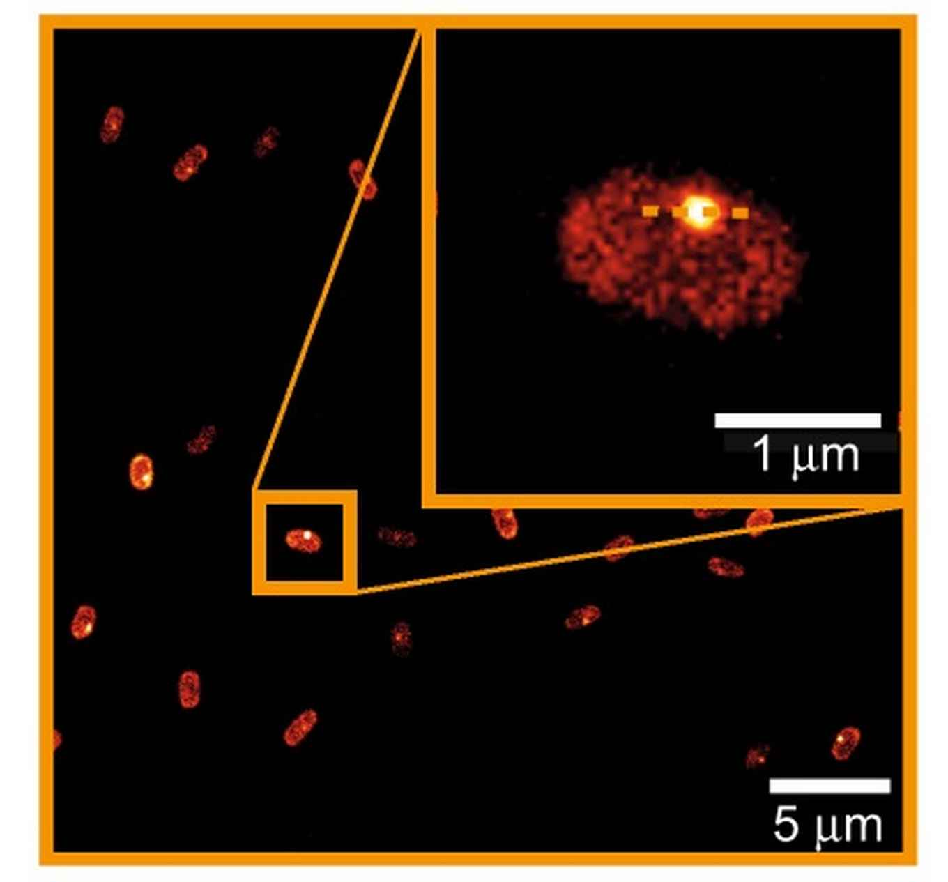
To prove the value of their technique, the team used it to study the germination of spores of the bacterium Bacillus subtilis. The germination, or ‘awakening’, of these spores is triggered by the binding of certain “germinants” to their receptor proteins (that can be fluorescently labelled) at so-called germinosomes. This process is notoriously hard to study, because the spores have very few of these germinosomes and because of autofluorescence of the spore coat. But the team’s new technique managed to overcome these problems, and they were able to perform time-lapse, super-resolution imaging of Bacillus subtilis spore germination.
Easy to use
According to the biologists themselves, their newly developed technique is architecturally less complex compared to other super-resolution microscopic techniques and easy to use. It uses standard preparation protocols and a relatively simple rescan confocal microscope with annular illumunation, and anyone with standard imaging expertise should be able to perform it. This means it could be adopted by any lab in need for higher resolution imaging.
Publication details:
R. M. P . Breedijk, J. Wen, V. Krishnaswami, T. Bernas, E. M. M. Manders, P. Setlow, N. O. E . Vischer & S. Brul; A live-cell super-resolution technique demonstrated by imaging germinosomes in wild-type bacterial spores, in: Nature Scientific Reports, 24 March 2020. DOI: https://doi.org/10.1038/s41598-020-62377-1