How do you uncover the innermost part of the brain?
17 April 2023
A hole in the brain
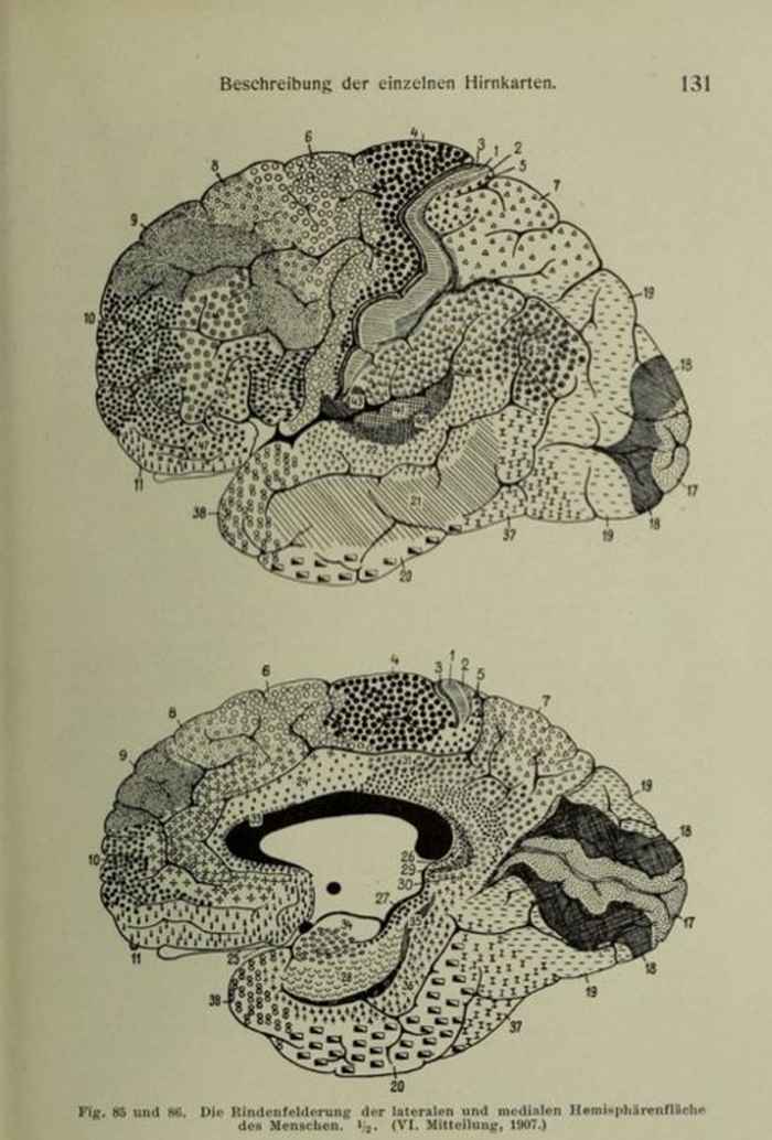
Mankind has been studying the brain for 2,000 years, making real headway in the last century in particular. This knowledge has been recorded in what might be termed ‘maps’ of the brain that designate its various areas and functions, such as the parts linked to sight and language.
‘All of those maps are really useful’, Miletić says, ‘and yet there’s something remarkable about them. That’s because they all show a blank area. It’s not like we have a hole in our brain, so why does every map have something missing from the centre?’
The subcortex
The missing part is known as the subcortex: an area immediately below the cortex, or the outer layer of the cerebrum. ‘There are various scientific clues that the subcortex is vital to healthy functioning’, Miletić says. ‘As an example, the subcortex is where the symptoms of Alzheimer’s and Parkinson’s disease appear first. It’s therefore essential to our understanding of brain functions and disease that we gain a thorough understanding of the subcortex.’ This leads us back to the question why this area is blank on all of the maps.
Difficult to measure
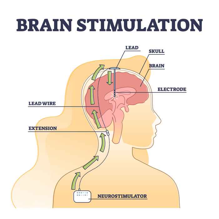
To find the answer, let’s take a look at the image on the right. ‘This is a good illustration of where the subcortex is located: at the centre of the brain, furthest away from the skull. It’s the area around the tip of the electrode. With live human subjects, we can only look inwards from the outside of the skull, for example by performing an MRI or EEG scan. However, the distance is too great to obtain a sharp image of the subcortex.’
How about looking inside the brain of a deceased subject? ‘Although it’s possible to obtain a clear image of the anatomy of a deceased subject, this isn’t the same as the anatomy of a healthy living person. The brain of a deceased subject has often aged dramatically. It may also show deformations and its shape changes when you remove it from the skull. Moreover, it’s impossible to detect which bodily functions or human behaviours are related to which parts of the brain when the brain is dead.’
Improved anatomic imaging with 7 Tesla
All this goes to show that the subcortex is a difficult area to research. Nevertheless, Miletić has succeeded in obtaining a better anatomic image using an MRI scanner with a powerful 7 Tesla magnet. ‘The most common MRI scanners are less powerful, resulting in a fuzzy grey image of the subcortex that hardly shows any detail at all. The 7 Tesla scanner makes it possible to detect more contrast in the tissue’, Miletić explains.
Operating the scanner correctly
While improved anatomic imaging is important, Miletić’s main interest as a neuroscientist was in finding out what links the subcortex to certain types of behaviour. This required him to map out the activity in this part of the brain. ‘For example, do certain areas light up when you display a particular behaviour, or do they remain inactive?’ In order to measure this, however, you must know how to operate the scanner correctly. ‘It’s not as simple as pushing a button. There’s a very long list of settings to choose from, and every choice you make affects the quality of the image.’ Consequently, the second step of his research focused on identifying the 7T scanner settings that would result in the best possible scan of the subcortex.
Performing tasks inside the scanner
Following completion of this step, Miletić asked human subjects to perform certain computer tasks while inside the scanner so that he could measure their brain activity. ‘We know from animal studies that choosing and learning behaviours are prominent activities in the subcortex, so we focused on these behaviours in our human subjects as well. We had them choose between various options, assigning different point values to each. This taught them which options were best.’ Miletić then analysed the outcomes using cognitive models that were capable of predicting some of the choices.
As a result, he made several discoveries about the anatomy of the subcortex and its role in choosing and learning behaviours. ‘This kind of “slow science” doesn’t lead to eye-popping results, but rather to small steps towards a better understanding.’
Thesis details
Steven Miletić, 2023, ‘Modelling structure and function of the human subcortex’. Supervisor: Prof. B.U. Forstmann. Co-supervisor: Dr P.L.E.A. Bazin. To read this doctoral thesis, search the UvA-DARE database, which lists all publications.
PhD thesis defence
Monday, 17 April, Miletić will defend his PhD thesis at 14:00 in the Agnietenkapel, Amsterdam. You can also watch this PhD defence ceremony here