New method developed for analysing 3D images of otoliths
9 June 2022
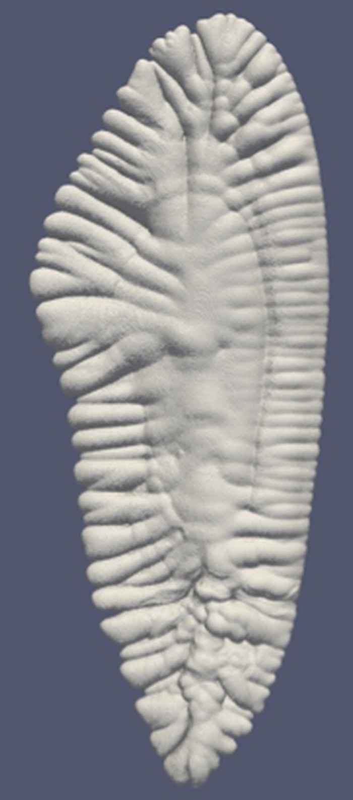
Images of the growth and form of otoliths in fish were obtained with micro Computed Tomography Scanning. This project was carried out in collaboration with the University of Bologna (Quinzia Palazzo).
Otoliths are intricate and complex shaped calcium carbonate structures in fishes which play a role in sound, movement and gravity detection. Growth rings in otoliths provide information about the age of the fish and analysis of otoliths is relevant in the population biology of fishes.
In this project, master student Steven Raaijmakers developed a method for quantifying these complex shaped morphologies as part of his master thesis work in Computational Science.
A paper about this work was recently accepted for publication in Royal Society Open Science .