UvA researchers establish record with exceptionally bright turquoise fluorescent protein
22 March 2012
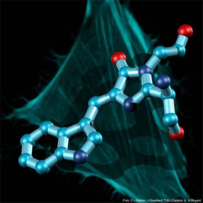
UvA researchers have succeeded in synthesising a turquoise-coloured fluorescent protein that is brighter than any other monomeric fluorescent protein. This feature makes it possible to more effectively study interactions between proteins in living cells. The researchers collaborated with Dr Antoine Royant’s group that is attached to the European Synchrotron Radiation Facility in Grenoble, France. Their findings were published on 20 March 2012 in Nature Communications.
The research shows that the efficiency with which the fully genetically encoded protein fluoresces (the quantum yield) is higher than any other monomeric fluorescent protein that has ever been measured. The synthesised protein has an efficiency of 93%, making the protein exceptionally fluorescent in living cells. Furthermore, the protein remains extraordinarily stable under the influence of light and is able to emit 1.6 million photons on average in a living cell before losing its luminosity. These properties make this protein very suitable for a wide range of applications in biomedical research, especially the study of disease-related processes in living organisms and cells.
Research methods
Molecular biologist Joachim Goedhart and colleagues from Prof. Dorus Gadella Jr.’s research group at the Swammerdam Institute for Life Sciences determined the structures of a number of cyan coloured (turquoise) fluorescent proteins. Based on an analysis of these protein structures, they were able to identify a number of amino acids that possibly had a negative influence on fluorescence. The DNA for these amino acids was subsequently randomly modified and transplanted in bacteria. These bacteria were then able to produce the corresponding fluorescent proteins.
The researchers combined this method with a recently developed technique for screening bacterial colonies for fluorescent protein variants created as a result of mutation. This process led to the isolation of exceptionally bright fluorescent turquoise protein referred to as ‘mTurquoise2’. Using X-ray diffraction, the researchers were able to establish the structure of ‘mTurquoise2’. They also studied the spectroscopic characteristics of the protein in vitro in cells.
The research was funded in part by Investment Subsidy NWO Medium, provided by the Netherlands Organisation for Scientific Research (NWO).
Publication information
J. Goedhart, D. von Stetten, M. Noirclerc-Savoye, M. Lelimousin, L. Joosen, M. A. Hink, L. van Weeren, Th.W.J. Gadella Jr. & A. Royant: ‘Structure-guided evolution of cyan fluorescent proteins towards a quantum yield of 93%’, in Nature Communications, 20 March 2012.