UvA scientists create highly detailed 3D reconstruction of a human brain
28 April 2022
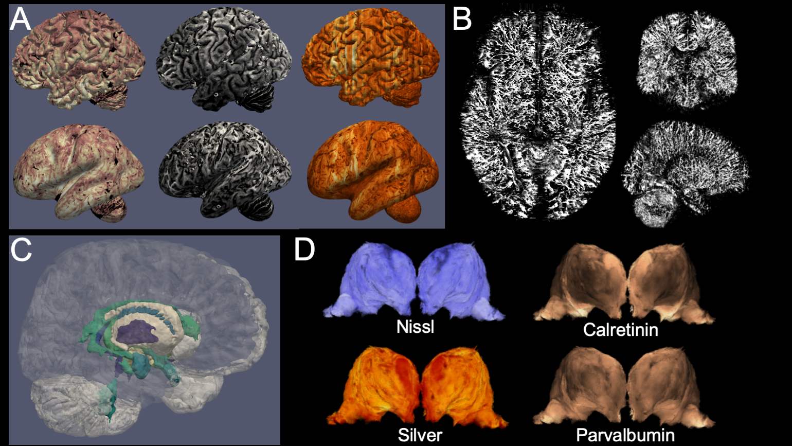
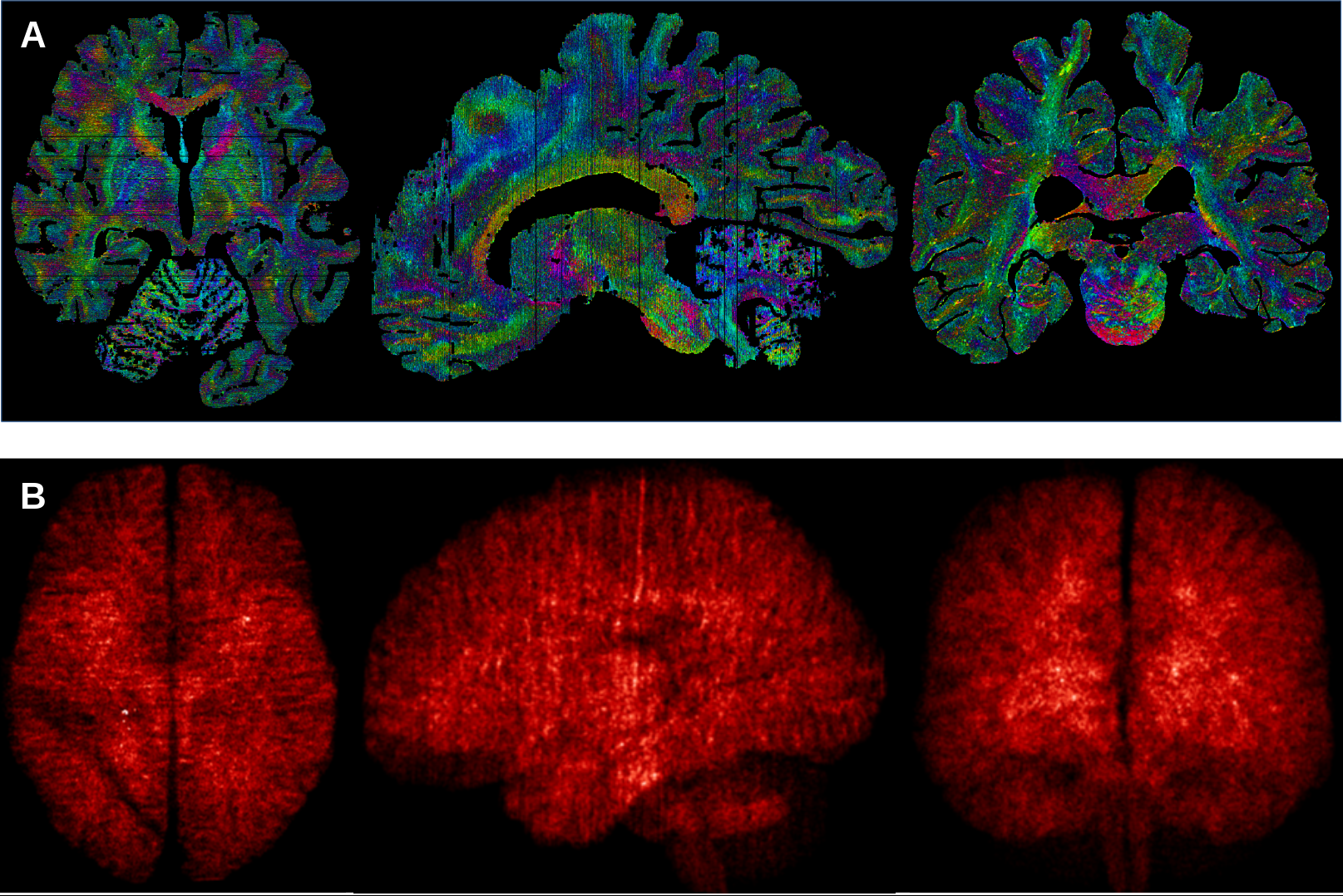
The UvA team worked for over five years, alongside researchers from the Max Planck Institute in Leipzig, to build a bridge between ultra-high field MRI and microscopy approaches to creating images of the brain. Two human brains donated to science were placed in the MRI scanner for 21 hours, and afterwards examined under the microscope. The MRIs were then combined with the microscopy slides, resulting in images of the brains which allow for exploration at a 200mm (0.2mm) level of detail.
Virtual brain dissections
Team member Anneke Alkemade: ‘We are excited about all the possibilities this can open up for the field. Instructors, for example, can use the datasets for neuroanatomy trainings or virtual dissections. And being able to compare MRI results with individual proteins visualised using microscopy will give researchers more insight into poorly understood MRI observations, as well as providing more anatomical detail on small brain structures.’
The researchers used an ultra-high field 7 Tesla MRI system, which has a more powerful magnet than the MRI systems routinely used in hospitals. The MRI software was programmed specifically for these studies by the researchers to accommodate the differences between living and preserved tissue. During the cutting of the tissue, each section was photographed individually, so that it could be used later to digitally correct tissue deformation in microscopy sections. Individual brain sections were placed on specially ordered glass slides, and processed with custom-built laboratory equipment.
After digitisation of the individual microscopy slides, new algorithms were created by the researchers to correct for the tissue deformation resulting from the cutting and microscopy processing. After weeks of uninterrupted calculations, the researchers were finally able to create full reconstructions of two individual brains.
Open science
Following Open Science principles, the researchers have made their data available free of cost as a service to the field. Scientists and other interested parties from around the world can now travel through the resulting 3D reconstructions of the human brain.
View the 3D reconstruction here
Publication details
Anneke Alkemade, Pierre-Louis Bazin, Rawien Balesar, Kerrin Pine, Evgeniya Kirilina, Harald Moller, Robert Trampel, Johan Kros, Max Keuken, Ronald Bleys, Dick Swaab, Andreas Herrler, Nikolaus Weiskopf and Birte Forstmann: ‘A unified 3D map of microscopic architecture and MRI of the human brain’, in: Science Advances (27 April 2022). DOI: 10.1126/sciadv.abj7892